Advanced 3T fMRI Neuroimaging for Precise Brain Mapping and Diagnosis
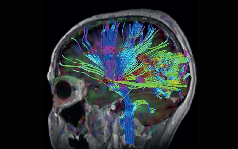
3T functional MRI (fMRI) uses a stronger magnet to produce detailed images of the brain, allowing for more precise diagnosis. The functional portion usually combined with a 3T brain MRI with and without contrast aids in detecting subtle changes in brain activity in certain neurological disorders or cognitive tasks. The scan sequences help neuroscientists discover how functional changes in the brain impact developmental and mental disorders including dyslexia, autism and schizophrenia.
A 3T fMRI is used for Neuroimaging to detect lesions, any abnormality or structural change in the brain and spinal cord. A 64-channel head coil provides better detail which aids in diagnosis. Functional brain mapping allows brain mapping during various cognitive and motor stimulation. This helps identify underlying conditions such as epilepsy, dementia, stroke, potentially Alzheimer’s.
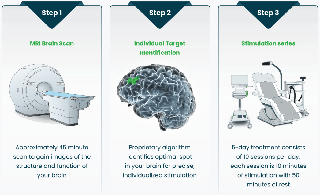
The 3T MRI has a higher sensitivity to detect subtle changes in blood oxygenation level-dependent (BOLD) contrast, which is a measure of brain activity. The 3T MRI also provides sharper resolution of images to localize brain activity. The data from the functional MRI portion is entered into a computer program called Localite. Localite analyzes the areas of the brain that need stimulation or destimulation based on the patient’s diagnosis. The patient’s brain can be treated within 1mm of the targeted area for transcranial magnetic stimulation (TMS).
The 3T MRI at Restorative Imaging is a wide bore which is larger and shorter, ideal for patients who struggle with claustrophobia. The cost is the same for a 1.5T MRI imaging procedure vs a 3T MRI imaging procedure. Patient time on the 3T scanner is sometimes less than a 1.5T MRI. The shorter time on the table minimizes the need for repeat sequences due to motion by the patient.
SAINT® Neuromodulation System
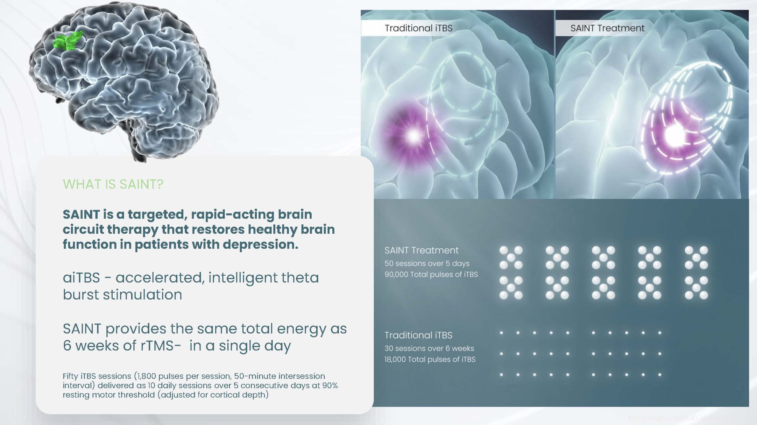
The Stanford Accelerated Intelligent Neuromodulation Treatment (SAINT®) system identifies the optimal therapy target in each person’s brain by using breakthrough algorithms with structural and functional MRI for precise, personalized treatment.
By precisely stimulating a person’s identified target with a specialized, non-invasive pattern of repetitive magnetic pulses, SAINT® effectively modifies activity in brain networks related to major depression and restores mood balance.
Restoring Healthy Brain Function
Depression is caused by identifiable changes in brain networks. SAINT® therapy restores neural networks, allowing individuals to feel more like themselves.
To learn more or request a consultation about the fMRI Guided TMS 5-Day Treatment visit restorativebraincenter.com.


